
News Release
These are the very first photomicrographs of nematode larval and adult worms and worm eggs in Alzheimer’s brain tissues with a tissue marker which is never found in normal brain tissues.
Cytokeratins are intermediate filaments produced in epithelial-derived cell lines. The brain has absolutely no cytokeratins in its healthy state.
Tumors which involve brain tissues are confirmed using immunostains for cytokeratin markers. Such tumors are chordomas, metastatic cancers, and pituitary tumors of the craniopharyngioma type.
Since parasitic worms are enveloped in epithelial (worm derived) tissues such as cuticle cells, it was reasoned that use of immunostains for epithelial cell lines ( Pan Cytokeratins) would demonstrate intact worms, worm fragments, and worm sloughed debris in parasitized brain tissues.
The use of a completely independent line of investigation ie Cytokeratin markers was determined to be a productive avenue for proof of the existence of nematode worms in the Alzheimer’s Disease autopsy brain.
Proof of nematode worm infestation in Alzheimer’s disease has never before been contemplated in the 100 years since Dr. Alois Alzheimer published his first case study of patient Auguste D..
Parasitic infestations of brain can be eradicated by appropriate treatment with anti-helminthic medications (analogous to the anti-parasitic medications which are routinely used in veterinary medical practice.)
Dr. Alan B. MacDonald, who has been researching and publishing evidence of the link between Alzheimer’s disease and Borrelia bacteria since the 1980’s, adds:
“The connection to the Nematode worms and Borrelia biofilms is as follows:
Borrelia ride as endosymbionts inside the bodies of parasitic Nematodes, which reside inside of Alzheimer’s disease brain.
When the worm dies, the endosymbiont Borrelia are released and these form Biofilm communities.
Borrelia biofilms are coated with antibacterial amyloids (as described by the Harvard Medical School group – Tanzi et al).
Alzheimer’s disease is actually a blended disease which combines Biofilm infections, intraneuronal Borrelia, Parasitic Nematodes, prozone phenomenon due to excess antigens released “as a comet-like storm” from liposomes of Borrelia and from sloughed proteins and structural constituents from the Nematode worms (Neural Larva Migrans-like Parasitosis).
Plural species of nematode worms are implicated in this infection/parasitosis cascade.”
(c) 2016, Copyright Alan B. MacDonald, MD, July 1, 2016, All rights reserved.
The superhighway to a cure of Alzheimer’s Disease is now at hand.
These revelations are the product of largely self-funded
research by Dr. Alan MacDonald MD, FCAP, director of the Dr. Paul H Duray Research Fellowship Endowment , Inc (an IRS Tax -Deductible 501c3b medical charity).
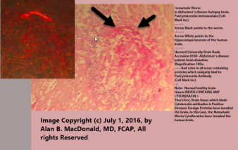
(Image at top right: Nematode worm in Alzheimer’s disease autopsy brain – Pan-Cytokeratin Immunostain (Cellmark Inc.) Black arrows point to the worm; white arrow indicates human hippocampal neurons. Red color in all areas contains proteins which uniquely bind to Pan Cytokeratin Antibody (Cellmark Inc.)
Note: Normal healthy brain tissues NEVER CONTAIN ANY CYTOKERATIN. The brain tissue which binds cytokeratin antibodies is positive in this image because foreign proteins are present in the human brain – in this case, due to invasion by nematode worms.
Source of tissue: Harvard University Brain Bank Accession 8100 – Alzheimer’s disease patient brain donation. Magnification 100x.
Image copyright July 1, 2016 by Dr. Alan MacDonald, MD, FCAP. All rights reserved.)
Website of Spirochaetal Alzheimer’s Association
Website of Dr Paul Duray Research Fellowship Foundation
Personal website of Alan B. MacDonald , MD, FCAP
Issued by: Spirochaetal Alzheimer’s Association
Email: alzheimer_borreliosis@yahoo.co.uk
